Left-Ventricular PVCs Improve Identification of Arrhythmic Risk in Mitral Valve Prolapse
Patients with mitral valve prolapse who have premature ventricular complexes originating from the left ventricle are at substantially increased risk of ventricular arrhythmias. The findings suggest that incorporating left-ventricular PVCs into the diagnostic criteria for arrhythmic mitral valve prolapse (AMVP), may improve identification of patients requiring closer surveillance.
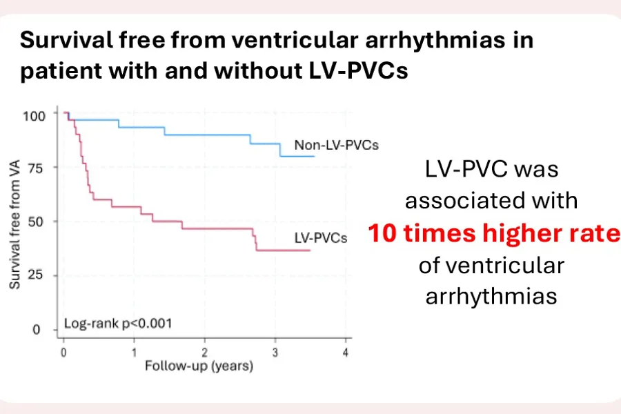
Figure: Adapted from Bugge C et al. Heart Rhythm O2. 2026. Article in press. DOI: https://doi.org/10.1016/j.hroo.2026.08.136
New publication by ProCardio
Left ventricular origin of arrhythmia predicts severe outcome in mitral valve prolapse: Insights from long-term cardiac monitoring (Bugge C et al. Heart Rhythm O2. 2026. Article in press).
Diagnostic criteria and risk stratification in patients with arrhythmic mitral valve prolapse (AMVP) are insufficient. The current AMVP diagnosis defined by EHRA includes burden of premature ventricular complexes (PVCs) but not the origin of PVCs. We explored left ventricular PVCs (LV-PVCs) as a risk factor for subsequent ventricular arrhythmia (VA) assessed by implantable cardiac monitor (ICM). We retrospectively applied the 2022 EHRA AMVP definition on our pre-2022 patient cohort.
In this prospective cohort study, we implanted ICM in patients with MVP and/or mitral annular disjunction during 2015-2020 and followed them for 3 years. We assessed occurrence of severe VA (ventricular fibrillation, sustained ventricular tachycardia [VT], or non-sustained VT [NSVT] causing syncope) and VA (severe VA and NSVT). PVCs origins were classified on 12-lead ECG.
We included 60 patients (mean age 49±15years) with ICM. LV-PVCs were present in 30 (50%) patients. LV-PVCs were associated with VA incidence rate ratio of 10.77 (95%CI 3.23-36.06, p<0.001), adjusted for baseline NSVTs and PVC burden. Severe VA occurred only in patients with LV-PVCs (log-rank p=0.04). The 2022 EHRA AMVP definition detected patients with subsequent VA with AUC 0.63, however, missed one (2%) patient with severe VA, and missed 12 (25%) patients with subsequent NSVT due to PVCs <5% at baseline.
Conclusion
Including presence of LV-PVCs in the AMVP diagnostic criteria and omitting the PVC threshold improved predictive accuracy and may help to correctly identify patients at arrhythmic risk for further surveillance. Further studies are needed to refine the AMVP definition and validate whether a lower PVC count and left-sided arrhythmia origin are potential factors to include in the definition of AMVP.